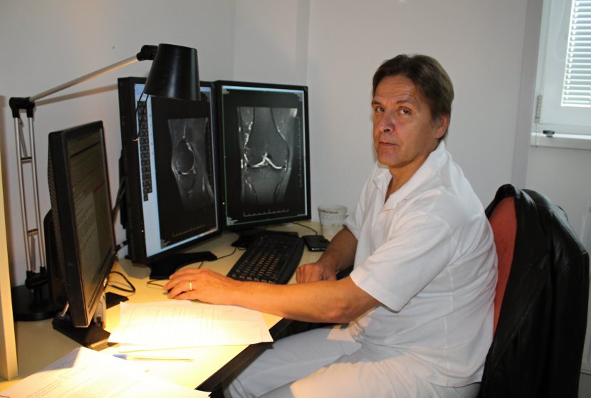
Doctor, could you briefly introduce yourself to our patients?
I have been working at the RN Beroun for 5 years as the head doctor of the Magnetic Resonance Imaging (MRI) department. I have years of experience with MRI, from 1995 to 2004 I worked at the MRI at the Radiodiagnostic Clinic of the 1st Faculty of Medicine of the Charles University in Prague, from 2005 to 2009 at the MRI at the Radiodiagnostic Clinic of the Bulovka Hospital, and from 2009 to 2012 as the head doctor of the MRI at the Outpatient Centre for Head and Neck Diseases.
How does RN Beroun compare with neighbouring hospitals in the field of imaging methods?
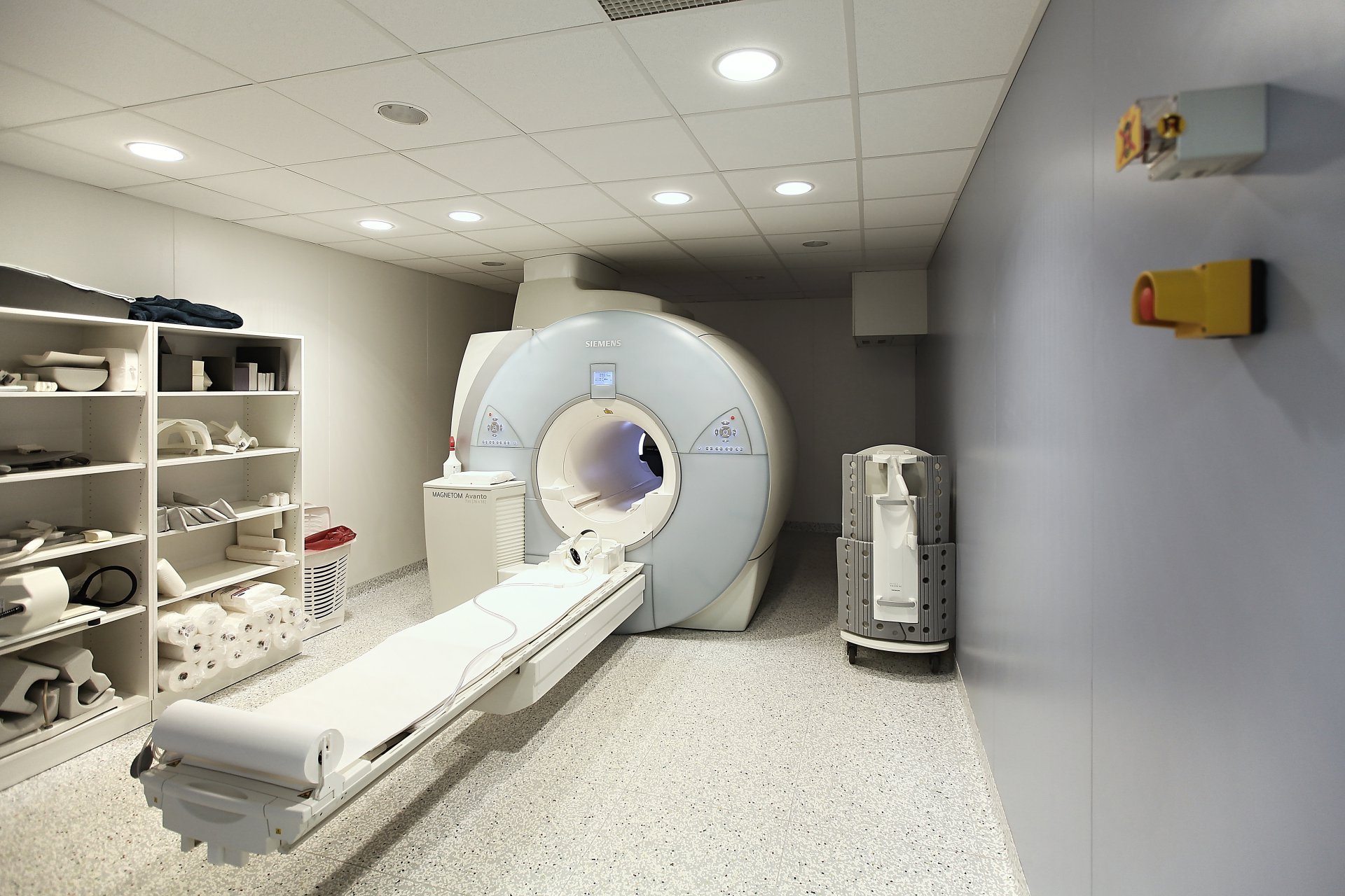
The Rehabilitation Hospital Beroun has the only MR machine within the hospital, where there is also a conventional X-ray and sonography. The disadvantage is that there is no CT scanner in RN Beroun, which is substituted by the hospital in Hořovice, for which we perform most MR examinations. Large hospitals in Prague have up to three MR machines, as is the case in Motol or VFN.
How many patients a year undergo a "magnet" examination in the Beroun hospital and how long are the waiting times for this examination?
Around 9,000 patients undergo MR examinations at our MRI scanner every year, and we examine up to 26 patients per day. The waiting time for an examination varies from 2 to 3 weeks, with demand depending significantly on the time of year. During the summer holidays, the waiting time is significantly shorter. The normal examination time for a patient is 30 minutes, but when examining multiple areas of interest, it can extend up to an hour for a single patient.
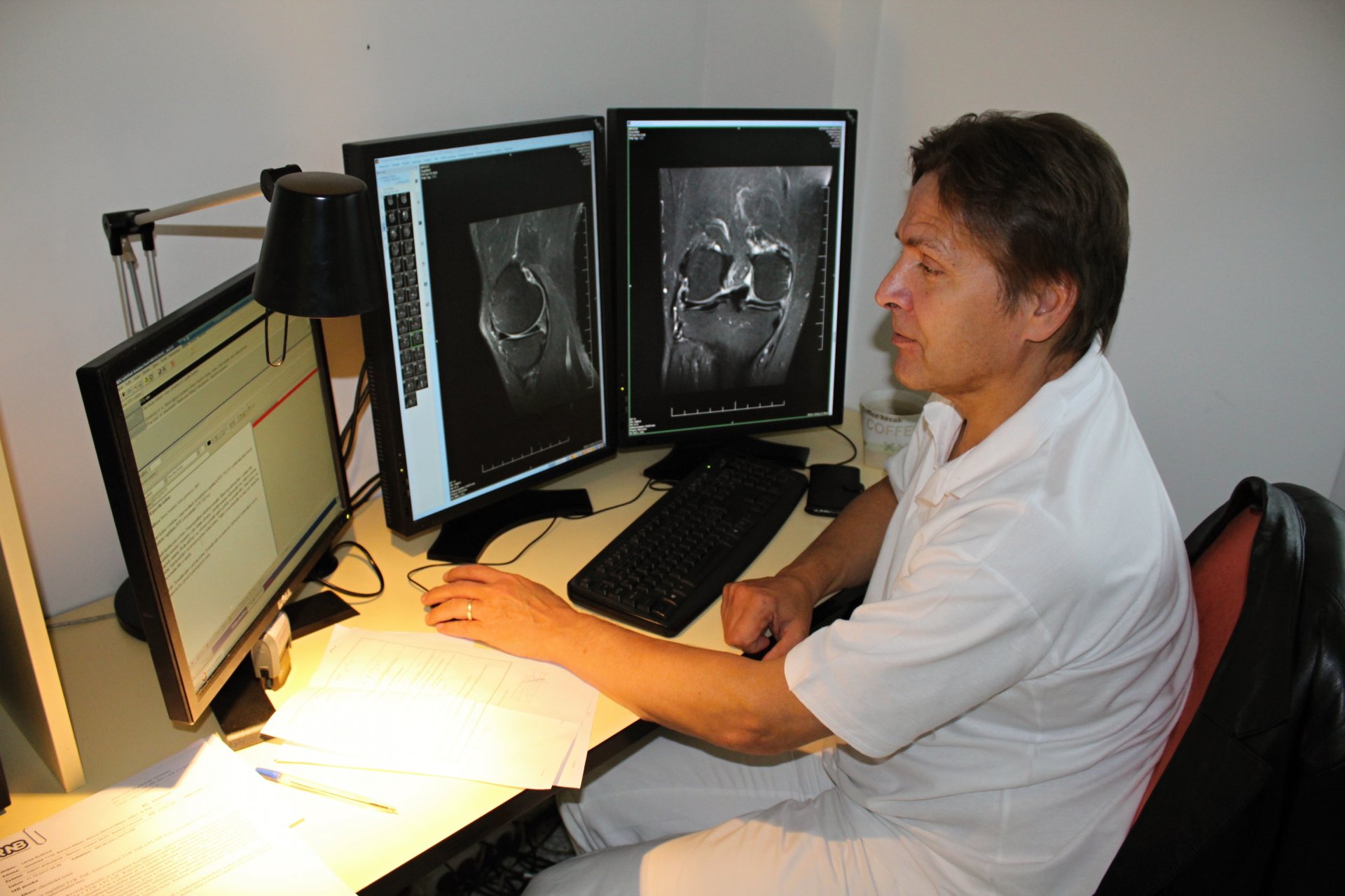
In which cases do patients undergo examinations using this imaging method and what can it reveal in the patient's body?
The MR examination is strictly targeted. We examine the brain, the cervical, thoracic or lumbar spine, individual large joints such as hips, shoulders, elbows, wrists or just individual fingers and their joints, if necessary. Unlike a CT scan, where we examine the entire abdomen clearly, with MRI it is necessary to target a specific organ such as the liver, kidneys, pancreas, spleen, rectum prostate etc. In most cases, MRI is performed as an adjunct to an already performed CT scan.
What preparation does this examination require, is it painful? Can all patients undergo it?
The MRI examination is painless, the disadvantage is the noise and the enclosed space of the tunnel into which the patient is introduced. We muffle the noise with "deafeners", which are similar to headphones for listening to music. The confined space is a problem in claustrophobic patients, where the fear of confined spaces can be medically influenced, in the extreme by general anaesthesia. This is only carried out by some centres as it requires special equipment operating under strong magnetic field conditions. Absolute contraindications for MR examinations are pacemakers and cochlear implants, which can endanger the patient's life by their presence during the MR examination. Relative contraindications are all metals in the patient's body. They must be present in the patient's body for more than 6 weeks, when any heating no longer interferes with tissue healing. For the examination of the liver, intestines and organs of the abdominal cavity, the patient must be fasting. Other examinations require no preparation. In some cases, it is necessary to administer a contrast agent intravenously during the examination. This is a different contrast from the iodine contrast used in CT. It is characterised by a significantly lower risk of allergic reaction. We just need to know the kidney function of the patient, when it fails, the excretion of contrast from the body is prolonged, which can be harmful to the patient in certain circumstances.
Is magnetic resonance imaging state-of-the-art? What developments are expected in the next few years in imaging methods?
MRI is the most advanced structural imaging method, complementary to other methods such as CT and conventional X-ray. These structural imaging methods are often combined with functional methods, which do not accurately depict structure but reliably reflect function. These methods include SPECT (Single Photon Emission Computerized Tomography) and PET (Positron Emission Tomography). The most sophisticated in this respect are hybrid devices where both structure and function are displayed in the same sections (PET-CT, PET-MR). These methods are used, for example, to search for secondary tumour deposits - metastases. In the field of MRI, the development is far from complete, the examination time is gradually shortening, new imaging modalities are being developed and are becoming more specific for individual diagnoses.


