Česky
Česky


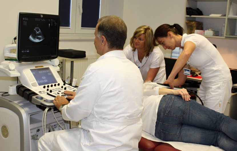
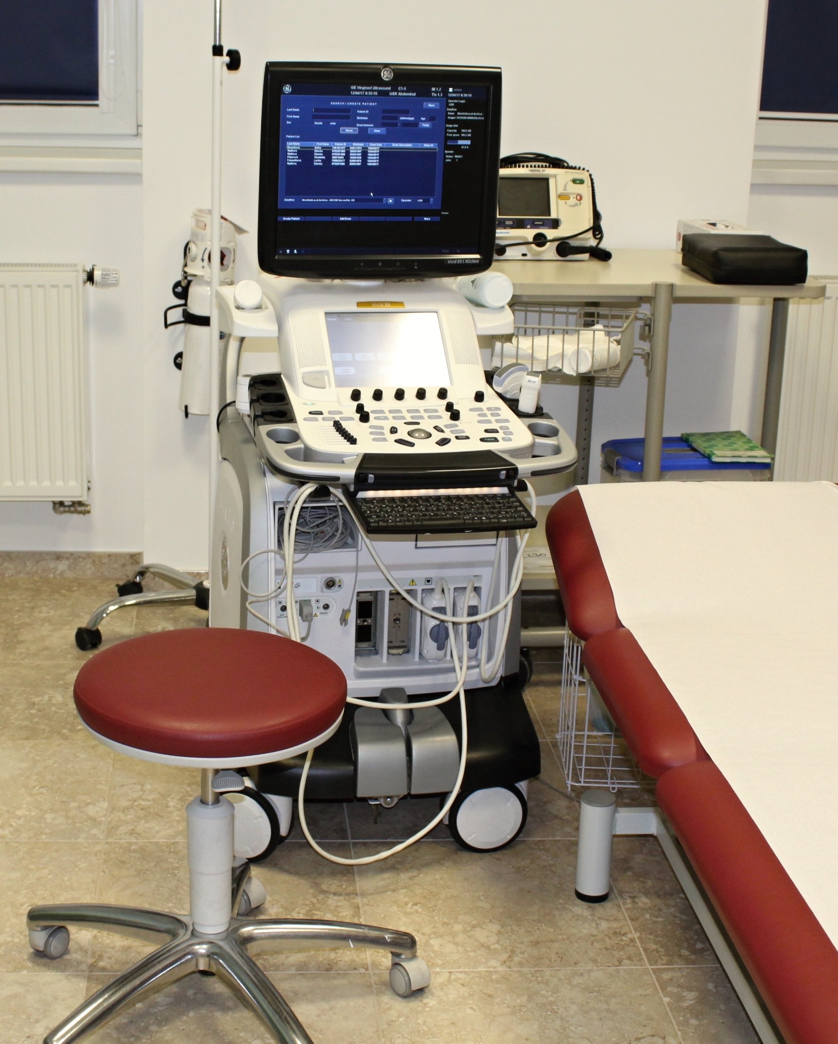
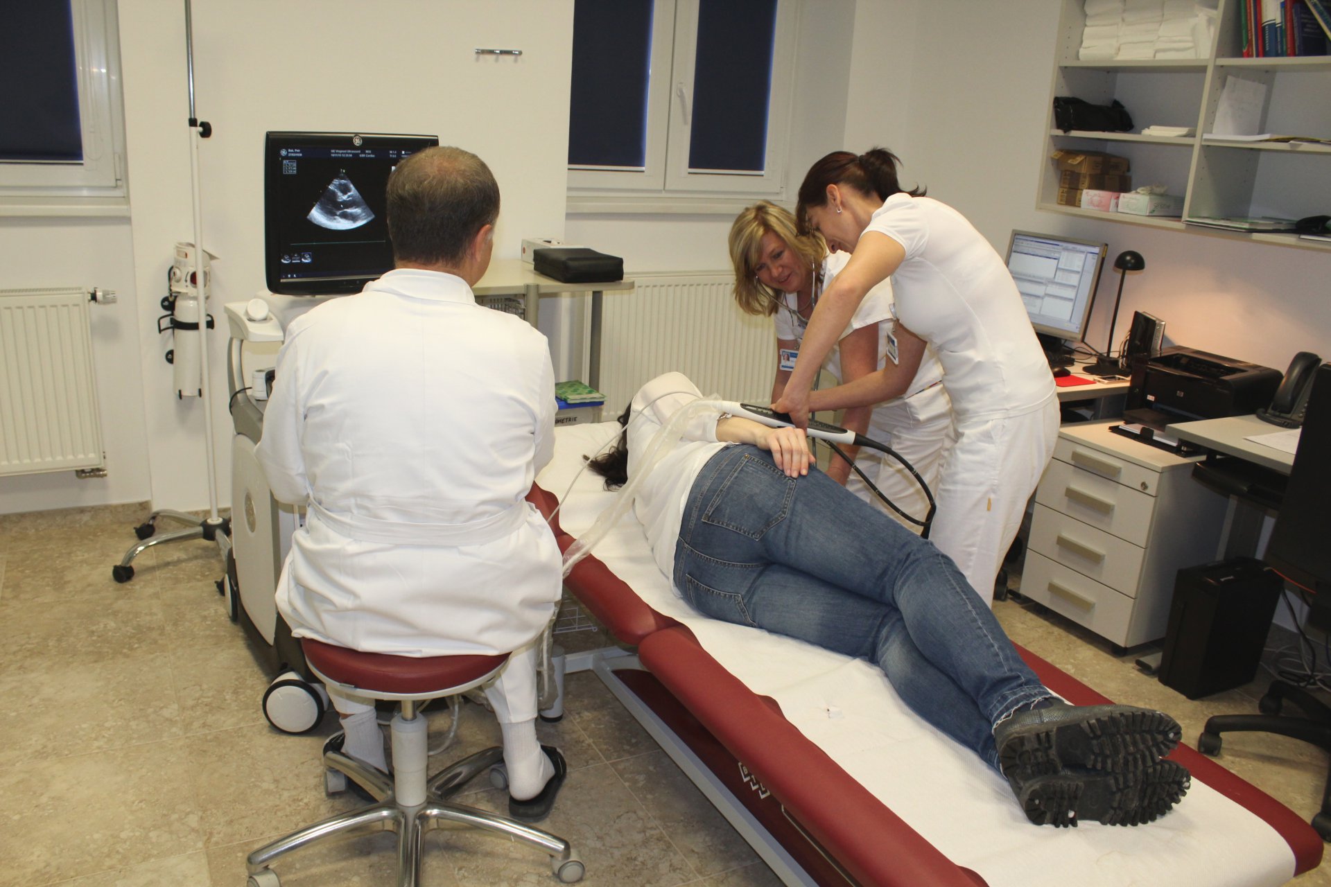
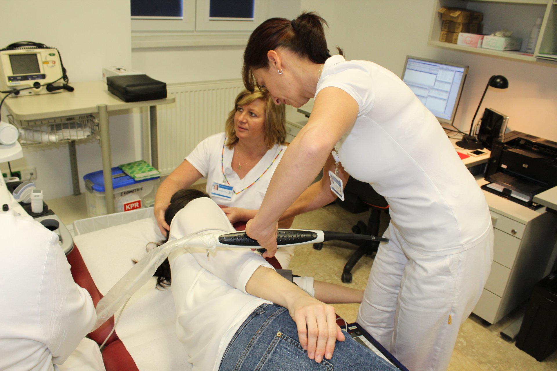
The basic principle of this examination is identical to the classical echocardiographic examination, the difference being that the ultrasound probe is not placed on the chest, but is placed at the end of an endoscope (a thin, steerable, flexible tube) that is introduced through the mouth into the oesophagus, to the point where the oesophagus runs adjacent to the heart. This gives a better and more detailed image than a conventional chest wall examination, and some special cardiac structures are not visible in the conventional way. "As an example, we can examine the so-called left atrial appendage, where blood clots often form, or search for infectious inflammation of the heart's intracardiac membranes on the heart valve or other intracardiac structures," says Jaromír Janoušek, MD, who together with Romana Zajacová, MD, and nurse Dagmar Bránecka, MD, performs this examination at the hospital in Beroun. He adds that this examination is also necessary during the preparation of the patient for the treatment of heart rhythm disorders, which are solved by electrical discharge, and during the complete examination of the patient after a stroke. We also asked whether the patient has any concerns about this examination and how it is performed. There is certainly no need for concern. The examination does not take long, during which basic vital signs are monitored, the patient is connected to an ECG, and his blood pressure is taken before and after. Even if it is necessary to inject a contrast agent into the vein, namely a concentrated glucose solution with microbubbles from which the ultrasound is well reflected, it is not burdensome and there is no risk of an allergic reaction. In order to prevent a gag reflex during the insertion of the probe, a numbing spray is applied to the pharynx; in addition, it is possible to administer intravenously not only anti-vomiting drugs, but also, and above all, soothing preparations.
All instructions for oesophageal echocardiography should be followed, the most important of which is to be fasting. After the examination, if the pharynx has been treated with a numbing spray, do not eat or drink for about 2 hours and, in the case of analgosedation, come to the examination with an escort and certainly do not drive or perform any activity that requires increased attention. The patient is always fully briefed on all details before and after the examination.