Česky
Česky


23.3.2011
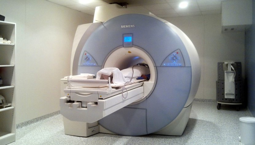
No more commuting and long waits for the necessary MRI scanSince March, the X-ray department of Beroun Hospital has been offering MRI examinations on a modern Siemens Magnetom Avanto 1.5 Tesla machine. Beroun Hospital is thus expanding its services in the imaging department by another qualitative leap. Among other things, by ensuring better availability of this modern diagnostic method for the residents of the local catchment area, but not only for them.
Magnetic resonance imaging (MRI), sometimes called "tunnel" among people, provides an examination that, like the perhaps better known "CT scan" for many, allows to show sections of the human body, but in the case of MRI without X-rays. This method has been used in practice for about 30 years, with its development preceded by a series of researches, bringing gradual steps towards its use in medicine. The ground-breaking discoveries were evidenced by the several Nobel Prizes awarded successively in 1937, 1991 and 2003 to several world-class chemists and physicists.This prize has only been awarded since 1901, otherwise it would certainly have been awarded to Nikola Tesla, who in 1882 made a major breakthrough in physics with the discovery of the rotating magnetic field. It is his surname that still denotes the unit of magnetic induction, which characterises the force effects of a magnetic field, and for the record 1.5 Tesla is about 30 000 times stronger than the Earth's magnetic field.
But back to the use of MRI in medicine. This examination is completely painless, definitely very gentle to the human body and also safe. It is an indispensable part of the complex of imaging methods used in modern medical science, providing doctors with important and accurate information about virtually all soft (not contrasting on X-rays, i.e. not clearly visible) tissues and organs in the body. This possibility is successfully used, for example, in the imaging of the brain and spinal cord, but also in the imaging of soft tissues of joints, such as ligaments or intervertebral discs, as well as blood vessels and so on. Magnetic resonance imaging offers precise localisation of problems that are difficult to detect by other imaging methods and many other diagnostic advantages. It also serves to prepare surgeons for complex reconstructive procedures, removal of soft tissue tumours, etc. It is true that there are some groups of patients for whom this examination cannot be used. These are mainly patients with implanted objects made of ferromagnetic materials, such as pacemakers, intracranial vascular clamps made of ferromagnetic material. In pregnant women in the first three months of pregnancy, given the ongoing organogenesis of the fetus, it is always advisable to consider the urgency and potential benefit of this examination and, if necessary, to postpone it until after the first trimester. The appropriateness of the MRI should be decided by the attending physician in consultation with MRI experts.