Česky
Česky


11.3.2010
You can make an appointment for this examination through the reception of the outpatient clinics by calling 311 745 272 or 311 745 329.Ultrasound examination can be performed only on the basis of a recommendation of your attending physician (a request for SONO/ECHO examination filled in by the referring physician).
More information about this examination and some interesting facts
Already at the end of the 18th century, the Italian biologist Spallanzani noticed, while studying the flight of bats, that they avoided all kinds of obstacles quite safely in the dark. In his book, written in 1794, he stated that this phenomenon is not related to visual perception, but to the previously unknown sound vibrations, inaudible to the human ear, which bats emit in flight, perceive after bouncing off obstacles and orient themselves according to them even in the most confined spaces.
The theoretical foundations of acoustics and ultra-acoustics were laid in 1877 by the English physicist Rayleigh. However, this discovery was not developed and exploited for quite a long time, the turning point came after the Titanic disaster, when a device for detecting icebergs by means of horizontally transmitted ultrasonic waves in the direction of the ship's sailing was soon patented in England. During the First World War the system was improved and used as an anti-submarine warning.
At the same time, similar devices began to be used in industry, especially for defectoscopy of materials, and the improvement of industrial reflexoscopes became the basis for the use of ultrasound in medicine.
Even before 1967, the first cardiological ultrasonograph was produced in the Škoda plant in Plzeň from an original industrial defectoscope. The impetus for it came from Prof. J. Sova, the then head of the cardiology-oriented Pilsen internal medicine clinic.
Medical ultrasound diagnostics is based on different reflection of ultrasound from different tissues. Echocardiography is a non-invasive (ultrasound) imaging method in cardiology whose rapid technological development over the previous 40 years has led to such a wide introduction into clinical practice that it actually represents one of the most significant diagnostic advances in medicine over the past century. Today, it is one of the most common examinations.
Instrumentation
The two instruments we use at Beroun Hospital are of a higher category and among the most modern on the market, and of course they undergo regular service checks. Doctors performing ultrasound examinations are professionally qualified.
- The Logic 9 sonography machine is a top-of-the-range sonograph with extensive capabilities, especially in the vascular area. LOGOQ 9 is the "flagship" of General Electric's ultrasound line, a high-performance Hi-End premium system for broad (shared services) use working with GE's revolutionary "TruScan Architecture" technology. The system offers unsurpassed performance, new imaging modes, excellent image data handling, flexible access and upgradeability.
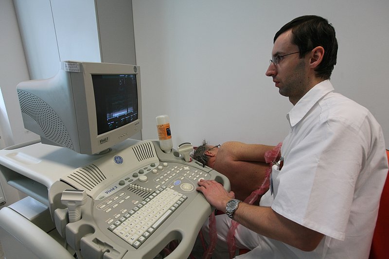
- The Vivid 3 enables complete highly specialised cardiac ultrasound with 1 and 2 dimensional echocardiography, pulsed and continuous Doppler, color blood flow mapping and higher harmonic component imaging. Together with computer processing of the results of the measurements taken, the device provides the most complete overview of cardiac activity in a non-invasive (non-bloody) manner.
Carotid sonography
There are two carotid arteries. Each runs on one side of the neck. Their job is to bring oxygenated blood to the brain. The carotid arteries branch off from the strongest artery in the body called the aorta. It comes out of the left ventricle of the heart. Carotid (carotid) ultrasonography images and functionally describes the blood flow through the carotid arteries. During the examination, the doctor determines whether these arteries are wide enough or narrow enough, at what speed and in what way the blood flows through them. This examination can also detect the early stages of cerebral anaemia and provide treatment or indicate the need for surgery. In simple terms, this examination can prevent ischaemic stroke or improve the condition of people who have already suffered a stroke.
For the patient, this examination is uncomplicated, completely painless, usually takes 5 to 10 minutes and no preparation is necessary beforehand. Your doctor will book you in for this examination if he or she suspects a poor blood supply to the brain. The first signals may be dizziness, headache, disturbances in thinking and consciousness. Patients with high blood pressure and high cholesterol are also screened as a preventive measure.
Echocardiography
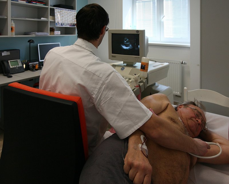
This is an ultrasound examination of the heart. Ultrasound is sent and received by an ultrasound probe that is placed on the chest. The ultrasound reflections from the heart are analysed by the machine and converted into a moving image of the heart walls and structures (e.g. heart valves). This allows the examination to show the individual heart compartments and valves, their structure, size and function. Modern devices also allow the direction and velocity of blood flow to be displayed, including an estimate of intracardiac pressures. This has made echocardiography of the heart an essential cardiac examination, replacing catheterisation in many cases.
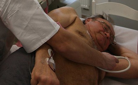
The patient lies on an examination couch, usually on the left side or supine. An ultrasound gel is applied to the probe and then the probe is placed at various points on the chest, with gentle pressure to ensure good contact with the wall. It is usually performed on the anterior chest wall on the left, at the lower edge of the ribs near the scaphoid process, or vice versa from the jugular fossa. Apart from slight pressure at the site, the examination is completely painless. A normal examination takes 10 to 30 minutes.
Ultrasound has no significant adverse effects on the body and therefore the echocardiographic examination can be repeated as required. No special preparation is necessary before the examination.
Our echocardiography outpatient clinic cooperates with the echocardiography department of the Na Homolce Hospital; doctors from this department perform examinations at the Beroun hospital twice a week. Since 1992, under the leadership of Prof. Niederle - the pioneer and founder of Czechoslovak echocardiography, the Echocardiography Laboratory of the Hospital Na Homolce has been a training centre for transthoracic (through the chest) and transesophageal (oesophageal) echocardiography, now under the supervision of Assoc. MUDr. Mandysová.
In case of need of complex superconciliary ECHO examination, we refer clients to this department with European echocardiography accreditation.