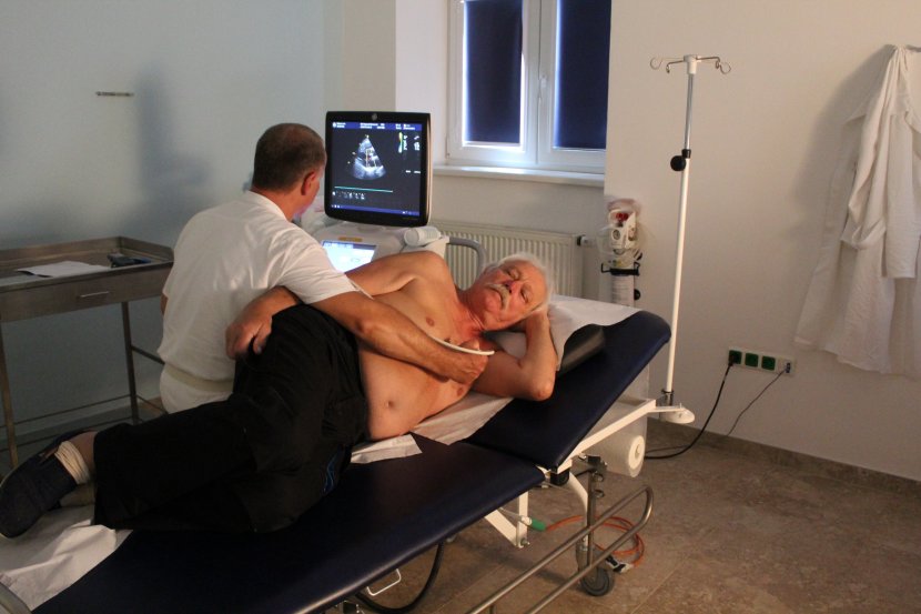
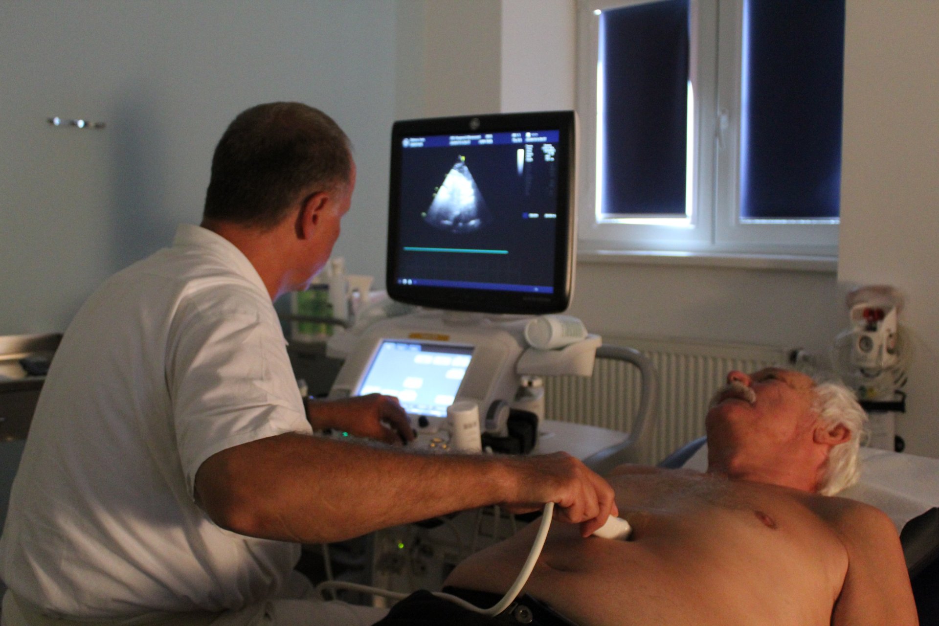
Echocardiography is an imaging method we use to perform an ultrasound examination of the heart. In our hospital, we use conventional transthoracic ECHO (examination through the chest wall) or transesophageal ECHO (examination through the esophagus).
Classical ECHO is a completely non-invasive, painless examination that requires no patient preparation. During the examination, the patient lies on the left side or on his/her back, and the ultrasound probe, which is placed on the patient's chest wall by the physician after applying the gel, is used to image the heart, its individual compartments and structures. This gives us information about the size of the heart, the size of its individual compartments, its ability to contract but also to relax, and whether any part of the heart is damaged, for example by poor blood supply. Furthermore, the doctor is interested in the thickness of the walls, the structure of the valves and their function (whether they are narrowed or, on the contrary, whether they do not tighten), and also measures the pressure gradients on the individual valves and sees whether there is, for example, fluid in the pericardium. Using this data, the doctor can obtain valuable information not only about morphological changes in the heart, but also about its function.
We asked Dr. Romana Zajacová what specific diseases the Echocardiograph is used for.
"We use ECHO to regularly monitor our internal medicine and cardiology patients - especially patients with coronary artery disease, arterial hypertension, various arrhythmias and cardiomyopathies, and patients after pulmonary embolisms and inflammatory heart diseases. As already mentioned, it is a painless, non-invasive and almost immediately available method (the only condition is the experience of the examining physician), so we can also use ECHO in acute situations - in the examination of patients with chest pain, shortness of breath, new arrhythmias, unconsciousness. We can thus more quickly determine the possible cause of the acute condition in a given patient and decide on further investigative and therapeutic procedures.
Esophageal ECHO is a more demanding examination, which already requires some preparation of the patient. The patient must be fasting. After local anaesthesia of the throat and usually after administration of medication to induce a slight attenuation and possibly reduction of the gag reflex, a probe is inserted into the oesophagus through the oral cavity. Since the oesophagus is in close proximity to the heart, we can view in much greater detail some structures that are harder to see or not at all in a conventional examination. We use this examination to diagnose infective endocarditis, detect the presence of intracardiac thrombi (clots) and shunts.
Through a pre-operative examination when the patient was going for hip replacement surgery, we discovered aortic valve narrowing through echocardiography, a serious diagnosis that ended up in a separate heart operation." The doctor further described the recovery.
Patients can make an appointment for the examination by referral from their treating physicians through our reception at 311 745 000. We currently screen every weekday during business hours and available appointments are available within one to two weeks.


